References
1. Foulks G, Bron AJ. A clinical description of meibomian gland dysfunction. Ocul Surf. 2003;1:107–126.
2. Kozak I, Bron AJ, Kucharova K, et al. Morphologic and volumetric studies of the meibomian glands in elderly human eyelids. Cornea. 2007;26:610–614.
3. Arita R, Itoh K, Inoue K, Amano S. Noncontact infrared meibography to document age-related changes of the Meibomian glands in a normal population. Ophthalmology. 2008;115:911–915.
4. Nicolaides N, Kaitaranta JK, Rawdah TN, Macy JI, Boswell FM 3rd, Smith RE. Meibomian gland studies: comparison of steer and human lipids. Invest Ophthalmol Vis Sci. 1981;20:522–536.
5. Chew CKS, Hykin PG, Jansweijer C, Dikstein S, Tiffany JM, Bron AJ. The casual level of meibomian lipids in humans. Curr Eye Res . 1993;12:255–259.
6. Yokoi N, Mossa F, Tiffany JM, Bron AJ. Assessment of meibomian gland function in dry eye using meibometry. Arch Ophthalmol. 1999;117:723–729.
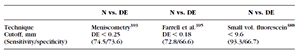
7. Yokoi N, Bron AJ, Tiffany J, et al. Reflective meniscometry: a non-invasive method to measure tear meniscus curvature. Br J Ophthalmol. 1999;83:92–97.
8. Chew CKS. Quantifying meibomian lipid on the human lid margin. Thesis. Oxford , UK : University of Oxford ; 1993.
9. Yokoi N, Bron AJ, Tiffany JM, et al. Relationship between tear volume and tear meniscus curvature. Arch Ophthalmol. 2004; 122:1265–1269.
10. Andrews JS. Human tear film lipids. I. Composition of the principal non-polar component. Exp Eye Res. 1970;10:223–227.
11. Nicolaides N, Ruth EC. Unusual fatty acids in the lipids of steer and human meibomian gland excreta. Curr Eye Res. 1982;2: 93–98.
12. Nicolaides N, Santos EC, Papadakis K, Ruth EC, Muller L. The occurrence of long chain alpha, omega-diols in the lipids of steer and human meibomian glands. Lipids. 1984;19:990–993.
13. Nicolaides N, Santos EC, Papadakis K. Double-bond patterns of fatty acids and alcohols in steer and human meibomian gland lipids. Lipids. 1984;19:264–277.
14. Nicolaides N, Santos EC. The di- and triesters of the lipids of steer and human meibomian glands. Lipids. 1985;20:454–467.
15. Tiffany JM. Individual variations in human meibomian lipid composition. Exp Eye Res. 1978;27:289–300.
16. Tiffany JM. The lipid secretion of the meibomian glands. Adv Lipid Res. 1987;22:1–62.
17. McCulley JP, Shine W. A compositional based model for the tear film lipid layer. Trans Am Ophthalmol Soc. 1997;95:79–88.
18. Sullivan BD, Evans JE, Dana MR, Sullivan DA. Influence of aging on the polar and neutral lipid profiles in human meibomian gland secretions. Arch Ophthalmol. 2006;124:1286–1292.
19. Butovich IA. Fatty acid composition of cholesteryl esters of human meibomian gland secretions. Steroids. 2010;75:726–733.
20. Butovich IA. The meibomian puzzle: Combining pieces together. Prog Retin Eye Res. 2009;28(6):483–498.
21. Butovich IA. Cholesteryl esters as a depot for very long chain fatty acids in human meibum. J Lipid Res. 2009;50:501–513.
22. Butovich IA. On the lipid composition of human meibum and tears: comparative analysis of nonpolar lipids. Invest Ophthalmol Vis Sci. 2008;49:3779–3789.
23. Butovich IA, Wojtowicz JC, Molai M. Human tear film and meibum: very long chain wax esters and (O-acyl)-omega-hydroxy fatty acids of meibum. J Lipid Res. 2009;50:2471–2485.
24. Butovich IA, Millar TJ, Ham BM. Understanding and analyzing meibomian lipids: a review. Curr Eye Res. 2008;33:405–420.
25. Butovich IA, Uchiyama E, McCulley JP. Lipids of human meibum: mass-spectrometric analysis and structural elucidation. J Lipid Res. 2007;48:2220–2235.
26. Nichols KK, Ham BM, Nichols JJ, Ziegler C, Green-Church KB. Identification of fatty acids and fatty acid amides in human meibomian gland secretions. Invest Ophthalmol Vis Sci. 2007; 48:34–39.
27. Linton RG, Curnow DH, Riley WJ. The Meibomian Glands: an investigation into the secretion and some aspects of the physiology. Br J Ophthalmol. 1961;45:718–723.
28. Shine WE, McCulley JP. The role of cholesterol in chronic blepharitis. Invest Ophthalmol Vis Sci. 1991;32:2272–2280.
29. Sullivan BD, Evans JE, Krenzer KL, Dana MR, Sullivan DA. Impact of antiandrogen treatment on the fatty acid profile of neutral lipids in human meibomian gland secretions. J Clin Endocrinol Metab. 2000;85:4866–4873.
30. Sullivan DA, Sullivan BD, Ullman MD. Androgen influence on the meibomian gland. Invest Ophthalmol Vis Sci. 2000;41:3732–3742.
Sullivan BD, Evans JE, Cermak JM, Krenzer KL, Dana MR, Sullivan DA. Complete androgen insensitivity syndrome: effect on human meibomian gland secretions. Arch Ophthalmol. 2002;120:1689–1699.
32. Sullivan DA, Sullivan BD, Evans JE, et al. Androgen deficiency, meibomian gland dysfunction, and evaporative dry eye. Ann NY Acad Sci. 2002:966:211–222.
33. Pes O. Ricerche microchimiche sulla secrezione delle ghiandole sebacee palpebrali. Arch Ottal 1897;5:82–91.
34. Ehlers N. The precorneal film. biomicroscopical, histological and chemical investigations. Acta Ophthalmol Suppl. 1965;(suppl)81: 81–134.
35. Cory CC, Hinks W, Burton JL, Shuster S. Meibomian gland secretion in the red eyes of rosacea. Br J Dermatol. 1973;89:25–27.
36. Keith CG. Seborrhoeic blepharo-kerato-conjunctivitis. Trans Ophthalmol Soc U K. 1967;87:85–103.
37. Harvey DJ, Tiffany JM, Duerden JM, Pandher KS , Mengher LS. Identification by combined gas chromatography-mass spectrometry of constituent long-chain fatty acids and alcohols from the meibomian glands of the rat and a comparison with human meibomian lipids. J Chromatogr. 1987;414:253–263.
38. Dougherty JM, McCulley JP. Analysis of the free fatty acid component of meibomian secretions in chronic blepharitis. Invest Ophthalmol Vis Sci. 1986;27:52–56.
39. Dougherty JM, McCulley JP, Silvany RE, Meyer DR. The role of tetracycline in chronic blepharitis. Inhibition of lipase production in staphylococci. Invest Ophthalmol Vis Sci. 1991;32:2970–2975.
40. Joffre C, Souchier M, Gregoire S, et al. Differences in meibomian fatty acid composition in patients with meibomian gland dysfunction and aqueous-deficient dry eye. Br J Ophthalmol. 2008;92: 116–119.
41. Joffre C, Souchier M, Leclere L, et al. Branched-chain fatty acids, increased in tears of blepharitis patients, are not toxic for conjunctival cells. Br J Ophthalmol. 2009;93(10):1391–1395.
42. Krenzer KL, Dana MR, Ullman MD, et al. Effect of androgen deficiency on the human meibomian gland and the ocular surface. J Clin Endocrinol Metab. 2000;85:4874–4882.
43. Mathers WD, Lane JA. Meibomian gland lipids, evaporation, and tear film stability. Adv Exp Med Biol. 1998;438:349–360.
44. Holly FJ. Formation and rupture of the tear film. Exp Eye Res. 1973;15:515–525.
45. Tiffany JM. Refractive index of meibomian and other lipids. Curr Eye Res. 1986;5(11):887–889.
46. Kocak I, Orgul S, Flammer J. Variability in the measurement of corneal temperature using a noncontact infrared thermometer. Ophthalmologica. 1999;213:345–349.
47. Bron AJ, Tiffany JM. The contribution of meibomian disease to dry eye. Ocul Surf. 2004;2(2):149–165.
48. Wolff E. Muco-cutaneous function of the lid margin and the distribution of the tear fluid. Trans Ophthalmol Soc UK . 1946; 66:291–308.
49. Blackie CA, Korb DR. Recovery time of an optimally secreting meibomian gland. Cornea. 2009;28:293–297.
50. Blackie CA, Korb DR . The diurnal secretory characteristics in individual meibomian glands. Cornea. 2009;29:34–38.
51. Norn M. Expressibility of meibomian secretion: relation to age, lipid precorneal film, scales, foam, hair and pigmentation. Acta Ophthalmol. 1987;65:137–142.
52. Korb DR, Blackie CA. Meibomian gland diagnostic expressibility: correlation with dry eye symptoms and gland location. Cornea. 2008;27(10):1142–1147.
53. Comberg W, Stoewer E. Die Augendruck-steingende Wirkung vershiedener: Muskelaktionen und ihre Bedeutun. Z Augenheilk. 1923;58:617.
54. Miller D. Pressure of the lid on the eye. Arch Ophthalmol. 1967;78:328–330.
55. Pflugfelder SC, Tseng S, Sanabria O, et al. Evaluation of subjective assessments and objective diagnostic tests for diagnosing tear-film disorders known to cause ocular irritation. Cornea. 1998;17:38–56.
56. Blackie CA, Korb DR, Knop E, Bedi R, Knop N, Holland EJ. Nonobvious obstructive meibomian gland dysfunction. Cornea . 2010;29:1333–1345.
57. Bron AJ, Tiffany JM, Gouveia SM, Yokoi N, Voon LW. Functional aspects of the tear film lipid layer. Exp Eye Res. 2004;78:347–360.
58. King-Smith PE , Hinel EA, Nichols JJ. Application of a novel interferometric method to investigate the relation between lipid layer thickness and tear film thinning. Invest Ophthalmol Vis Sci. 2010;51(5):2418–2423.
59. King-Smith PE, Fink BA, Nichols JJ, Nichols KK, Braun RJ, McFadden GB. The contribution of lipid layer movement to tear film thinning and breakup. Invest Ophthalmol Vis Sci. 2009; 50(6):2747–2756.
60. Owens H, Phillips JR. Spreading of tears after a blink: velocity and stabilization time in healthy eyes. Cornea. 2001;20:484–487.
61. Owens H, Phillips JR. Tear spreading rates: post-blink. Adv Exp Med Biol. 2002;506:1201–1204.
62. Goto E, Tseng SC. Differentiation of lipid tear deficiency dry eye by kinetic analysis of tear interference images. Arch Ophthalmol. 2003;121:173–180.
63. Doane MG. Interactions of eyelids and tears in corneal wetting and dynamics of the normal human eyeblink. Am J Ophthalmol. 1980;89:507–516.
64. Tsubota K. Tear dynamics and dry eye. Prog Retin Eye Res. 1998;17:565–596.
65. McCulley JP, Shine WE. Meibomian gland and tear film lipids: structure, function and control. Adv Exp Med Biol. 2002;506: 373–378.
66. Goto E, Tseng SC. Kinetic analysis of tear interference images in aqueous tear deficiency dry eye before and after punctal occlusion. Invest Ophthalmol Vis Sci. 2003;44:1897–1905.
67. Rolando M, Valente C, Barabino S. New test to quantify lipi layer behavior in healthy subjects and patients with keratoconjunctivitis sicca. Cornea. 2008;27(8):866–870.
68. McDonald JE. Surface phenomena of the tear film. Am J Ophthalmol. 1969;67:56–64.
69. DEWS. Methodologies to Diagnose and Monitor Dry Eye Disease: Report of the Diagnostic Methodology Subcommittee of the International Dry Eye WorkShop. Ocul Surf. 2007;5:108–152.
70. Behrens A, Doyle JJ, Stern L, et al. Dysfunctional tear syndrome: a Delphi approach to treatment recommendations. Cornea. 2006; 25:900–907.
71. Duke-Elder WS, MacFaul PA. The ocular adnexa, Part I: inflammations of the lid margins. Vol 13. In: System of Ophthalmology . London : H. Kimpton. 1974:205–250.
72. McCulley JP, Sciallis GF. Meibomian keratoconjunctivitis. Am J Ophthalmol. 1977;84(6):788–793.
73. Gutgesell VJ, Stern GA , Hood CI. Histopathology of meibomian gland dysfunction. Am J Ophthalmol. 1982;94:383–387.
74. Luchs J. Efficacy of topical azithromycin ophthalmic solution 1% in the treatment of posterior blepharitis. Adv Ther. 2008;25(9): 858–870.
75. Bron AJ, Benjamin L, Snibson GR. Meibomian gland disease. classification and grading of lid changes. Eye. 1991;5:395–411.
76. Schrader S, Mircheff AK , Geerling G. Animal models of dry eye. Dev Ophthalmol. 2008;41:298–312.
77. Bron AJ, Tiffany JM. The evolution of lid margin changes in blepharitis. In: Lass JH, ed. Advances in Corneal Research: Selected Transactions of the World Congress on the Cornea IV . New York : Plenum Press: 1997:3–18.
78. Obata H. Anatomy and histopathology of human meibomian gland. Cornea. 2002;21(7 Suppl):S70–S74.
79. Matsumoto Y, Sato EA, Ibrahim OM , et al. The application of confocal microscopy to the diagnosis and evaluation of meibomian gland dysfunction. Mol Vis. 2008;14:1263–1271.
80. Korb DR , Henriquez AS. Meibomian gland dysfunction and contact lens intolerance. J Am Optom Assoc. 1980;51(3):243–251.
81. Korb DR, Blackie CA. Diagnostic versus therapeutic meibomian gland expression (abstract). American Academy of Optometry annual meeting, Orlando , 2009. Available at http://www.aaopt.org/Submission/Search/ SubmissionViewer.asp?SID = 25745&BR = SP. Abstract 90745.
Goto E, Shimazaki J, Monden Y, et al. Low-concentration homogenized castor oil eye drops for noninflamed obstructive meibomian gland dysfunction. Ophthalmology. 2002;109(11):
2030–2035.
83. Hykin PG, Bron AJ. Age-related morphological changes in lid margin and meibomian gland anatomy. Cornea. 1992;11:334–342.
84. Lemp MA. Report of the National Eye Institute/Industry Workshop on Clinical Trials in Dry Eyes. CLAO J. 1995;21:221–231.
85. DEWS. The definition and classification of dry eye disease: report of the Definition and Classification Subcommittee of the International Dry Eye WorkShop. Ocul Surf. 2007;5:75–92.
86. Shimazaki J, Sakata M, Tsubota K. Ocular surface changes and discomfort in patients with meibomian gland dysfunction. Arch Ophthalmol. 1995;113:1266–1270.
87. Shimazaki J, Goto E, Ono M, et al. Meibomian gland dysfunction in patients with Sjögren syndrome. Ophthalmology. 1998;105: 1485–1488.
88. Driver PJ, Lemp MA. Meibomian gland dysfunction. Surv Ophthalmol. 1996;40(5):343–367.
89. Lemp MA, Nichols KK. Blepharitis in the United States 2009: a survey-based perspective on prevalence and treatment. Ocul Surf. 2009;7(suppl 2):S1–S14.
90. Mathers WD. Ocular evaporation in meibomian gland dysfunction and dry eye. Ophthalmology. 1993;100:347–351.
91. Mathers WD. Meibomian gland disease. In: Pflugfelder SC , Stern ME, Beuerman RW, eds. Dry Eye and the Ocular Surface: A Unified Approach. New York : Marcel Dekker. 2004:chap 22.
92. Khanal S, Tomlinson A, McFadyen A, Diaper C, Ramaesh K. Dry eye diagnosis. Invest Ophthalmol Vis Sci. 2008;49:1407–1414.
93. Tomlinson A, Doane MG, McFadyen A. Inputs and outputs of the lacrimal system: review of production and evaporative loss. Ocul Surf. 2009;7(4):186–198.
94. Robin JB, Jester JV, Nobe J, Nicolaides N, Smith RE. In vivo transillumination biomicroscopy and photography of meibomian gland dysfunction; a clinical study. Ophthalmology. 1985;92: 1423–1426.
95. Ong BL, Hodson SA, Wigham T, Miller F, Larke JB. Evidence for keratin proteins in normal and abnormal human meibomian fluids. Curr Eye Res. 1991;10:1113–1119.
96. Mathers WD, Shields WJ, Sachdev MA, Petroll WM, Jester JV. Meibomian gland dysfunction in chronic blepharitis. Cornea. 1991;10:277–285.
97. Mathers WD, Shields WJ, Sachdev MS, et al. Meibomian gland morphology and tear osmolarity: changes with Accutane therapy. Cornea. 1991;10:286–290.
98. Henriquez AS, Korb DR. Meibomian glands and contact lens wear. Br J Ophthalmol. 1981;65:108–111.
99. Yamaguchi M, Kutsuna M, Uno T, et al. Marx line: fluorescein staining line on the inner lid as indicator of meibomian gland function. Am J Ophthalmol. 2006;141:669–675.
100. Marx E. Uber vitale Farbung des Auges und der Augenlieder. I. Uber Anatomie, Physiologie und Pathologie des Augenlidrandes under der Tranenpunkte. Graefes Arch Clin Exp Ophthalmol. 1924;114:465–482.
101. Marx E. Uber vitale Farbung des Auges und der Augenlieder. I. Uber Anatomie, Physiologie und Pathologie des Augenlidrandes under der Tranenpunkte. Graefes Arch Clin Exp Ophthalmol. 1926;116:114–125.
102. Doughty MJ, Naase T, Donald C, Hamilton L, Button NF. Visualization of “Marx's line” along the marginal eyelid conjunctiva of human subjects with lissamine green dye. Ophthalmic Physiol Opt. 2004;24:1–7.
103. Hughes C, Hamilton L, Doughty MJ. A quantitative assessment of the location and width of Marx's line along the marginal zone of the human eyelid. Optom Vis Sci. 2003;80:564–572.
104. DEWS. Meibography/Meiboscopy. Template 25. 2007c. Available online only at: http://www.tearfilm.org/dewsreport/pdfs/
MeibographyMeiboscopy%20(Foulks).pdf Accessed Sept. 29, 2010.
105. Jester JV, Nicolaides N, Smith RE. Meibomian gland dysfunction, I: keratin protein expression in normal human and rabbit meibomian glands. Invest Ophthalmol Vis Sci. 1989;30:927–935
106. de Paiva CS, Lindsey JL, Pflugfelder SC. Assessing the severity of keratitis sicca with videokeratoscopic indices. Ophthalmology . 2003;110:1102–1109.
107. Nichols JJ, Berntsen DA, Mitchell GL, Nichols KK. An assessment of grading scales for meibography images. Cornea. 2005;24:382–388.
108. Mathers WD, Billborough M. Meibomian gland function and giant papillary conjunctivitis. Am J Ophthalmol. 1992;114:188–192.
109. Arita R, Itoh K, Inoue K, Kuchiba A, Yamaguchi T, Amano S. Contact lens wear is associated with decrease of meibomian glands. Ophthalmology. 2009;116(3):379–384.
110. Bailey IL, Bullimore MA, Raasch TW, Taylor HR. Clinical grading and the effects of scaling. Invest Ophthalmol Vis Sci. 1991;32: 422–432.
111. Efron N, Morgan PB, Katsara SS. Validation of grading scales for contact lens complications. Ophthalmic Physiol Opt. 2001;21: 17–29.
112. Paugh JR, Marsden HJ, Edrington TB, DeLand PN, Simmons PA, Vehige JG. A pre-application drop containing carboxymethylcellulose can reduce multipurpose solution-induced corneal staining. Optom Vis Sci. 2007;84:65–71.
113. Sparrow NA, Frost NA, Pantelides EP, Laidlaw DA. Decimalization of The Oxford Clinical Cataract Classification and Grading System. Ophthalmic Epidemiol. 2000;7:49–60.
114. McCann LC, Tomlinson A, Pearce EI, Diaper C. Tear and meibomian gland function in blepharitis and normals. Eye Contact Lens. 2009;35(4):203–208.
115. Mathers WD, Lane JA, Sutphin JE, et al. Model for ocular tear film function. Cornea. 1996;15:110–119.
116. Hykin PG, Bron AJ. Age-related morphological changes in lid margin and meibomian gland anatomy. Cornea. 1992;11:334–342.
117. Bron AJ, Yokoi N, Gaffney E, Tiffany JM. Predicted phenotypes of dry eye: proposed consequences of its natural history. Ocul Surf. 2009;7:78–92.
118. Khanal S, Tomlinson A, Diaper CJ. Tear physiology of aqueous deficiency and evaporative dry eye. Optom Vis Sci. 2009;86(11): 1235–1240.
119. Suzuki T, Mitsuishi Y, Sano Y, Yokoi N, Kinoshita S. Phlyctenular keratitis associated with meibomitis in young patients. Am J Ophthalmol. 2005;140(1):77–82.
120. Lam H, Bleiden L, de Paiva CS, Farley W, Stern ME, Pflugfelder SC. Tear cytokine profiles in dysfunctional tear syndrome. Am J Ophthalmol. 2009;147(2):198–205-e1.
121. Goto E, Endo K, Suzuki A, et al. Tear evaporation dynamics in normal subjects and subjects with obstructive meibomian gland dysfunction. Invest Ophthalmol Vis Sci. 2003;44:533–539.
122. Yokoi N. [Tear dynamics and dry eye.] Nippon Ganka Gakkai Zasshi. 2004;108:275–276.
123. Yokoi N, Yamada H, Mizukusa Y, et al. Rheology of tear film lipid layer spread in normal and aqueous tear-deficient dry eyes. Invest Ophthalmol Vis Sci. 2008;49:5319–5324.
124. Yokoi N, Komuro A. Non-invasive methods of assessing the tear film. Exp Eye Res. 2004;78(3):399–407.
125. McCulley JP, Dougherty JM, Deneau DG. Classification of chronic blepharitis. Ophthalmology. 1982;89(10),1173–1180.
126. Martin NF, Rubinfeld RS, Malley JD, Manzitti JD. Giant papillary conjunctivitis and meibomian gland dysfunction blepharitis. CLAO J. 1992;18:165–169.
127. Ong BL, Larke JR. Meibomian gland dysfunction: some clinical, biochemical and physical observations. Ophthalmic Physiol Opt. 1990;10:144–148.
128. Huber-Spitzy V, Baumgartner I, Böhler-Sommeregger K, Grabner G. Blepharitis—a diagnostic and therapeutic challenge. A report on 407 consecutive cases. Graefes Arch Clin Exp Ophthalmol. 1991;229:224–227.
129. Huber-Spitzy V, Böhler-Sommeregger K, Arocker-Mettinger E, Grabner G. Ulcerative blepharitis in atopic patients—is Candida species the causative agent? Br J Ophthalmol. 1992;76(5):272–274.
Blackman HJ, Recka GL, Olsen TG, Bergsma DR , Blepharoconjunctivitis: a side effect of 13-cis-retinoic acid therapy for dermatologic diseases. Ophthalmology. 1979;86:753–759.
131. Fraunfelder FT, LaBraico JM, Meyer SM. Adverse ocular reactions possibly associated with isotretinoin. Am J Ophthalmol. 1985; 100:534–537.
132. McMonnies CW. Key questions in a dry eye history. J Am Optometric Assoc. 1986;57:512–517.
133. Johnson ME, Murphy PJ. Measurement of ocular surface irritation on a linear interval scale with the ocular comfort index. Invest Ophthalmol Vis Sci. 2007;48(10):4451–4458.
134. Mengher LS, Bron AJ, Tonge SR, et al. Effect of fluorescein instillation on the precorneal tear film stability. Curr Eye Res. 1985;4:9–12.
135. Sakamoto R, Bennett ES, Henry VA, et al. The phenol red thread tear test: a cross-cultural study. Invest Ophthalmol Vis Sci. 1993; 34:3510–3514.
136. Patel S, Farrell J, Blades KJ, Grierson DJ. The value of a phenol red impregnated thread for differentiating between the aqueous and non-aqueous deficient dry eye. Ophthalmic Physiol Opt. 1998; 18:471–476.
137. van Bijsterveld OP. Diagnostic tests in the Sicca syndrome. Arch Ophthalmol. 1969;82(1):10–14.
138. Vitali C, Bombardieri S, Johsson R, et al. Classification criteria for Sjogren's syndrome: a revised version of the European criteria proposed by the American-European Consensus Group. Ann Rheum Dis . 2002;61(6):554–558.
139. Kobayashi A, Yoshita T, Sugiyama K. In vivo findings of the bulbar/palpebral conjunctiva and presumed meibomian glands by laser scanning confocal microscopy. Cornea. 2005;24(8):985–988.
140. Matsumoto Y, Dogru M, Sato EA, et al. The application of in vivo confocal scanning laser microscopy in the management of Acanthamoeba keratitis. Mol Vis. 2007;13:1319–1326.
141. Farrell J, Patel S, Grierson DG, Sturrock RD. A clinical procedure to predict the value of temporary occlusion therapy in keratoconjunctivitis sicca. Ophthalmic Physiol Opt. 2003;23:1–8.
142. Mainstone JC, Bruce AS, Golding TR. Tear meniscus measurement in the diagnosis of dry eye. Curr Eye Res. 1996;15:653–661.
143. Xu KP, Yagi Y, Toda I, Tsubota K. Tear function index. a new measure of dry eye. Arch Ophthalmol. 1995;113:84–88.
144. McCann LC, Tomlinson A, Pearce EI, Khanal S, Kaye SB, Fisher AC. A clinical alternative to fluorophotometry for measuring tear production in the diagnosis of dry eye. Cornea. 2010;29(7):745–750.
145. Ousler GW 3rd, Hagberg KW, Schindelar M, Welch D, Abelson MB. The Ocular Protection Index. Cornea. 2008;27(5):509–513.
146. Schiffman RM, Christianson MD, Jacobsen G, Hirsch JD, Reis BL. Reliability and validity of the Ocular Surface Disease Index. Arch Ophthalmol. 2000;118(5):615–621.
147. Begley CG, Caffery B, Nichols K, Mitchell GL, Chalmers R; DREI study group: results of a dry eye questionnaire from optometric practices in North America . Adv Exp Med Biol. 2002;506:1009–1016.
148. McMonnies CW. Key questions in a dry eye history. J Am Optom Assoc , 1986:57;512–517.
149. Schiffman RM, Christianson MD, Jacobsen G, Hirsch JD, Reis BL. Reliability and validity of the Ocular Surface Disease Index. Arch Ophthalmol. 2000;118(5):615–621.
150. Bandeen Roche R, Mun˜oz B, Teilsch JM, West SK, Schein OD. Self-reported assessment of dry eye in a population-based setting. Invest Ophthalmol Vis Sci. , 1997;38(12):2469–2475.
151. Johnson , ME , Murphy PJ. Measurement of ocular surface irritation on a linear interval scale with the ocular comfort index. Invest Ophthalmol Vis Sci. , 2007;48(10):4451–4458.
152. Begley CG, Caffery B, Chalmers RL, Mitchell GL; Dry Eye Investigation (DREI) Study Group. Use of the dry eye questionnaire to measure symptoms of ocular irritation in patients with aqueous tear deficient dry eye. Cornea. 2002;21(7):664–670.
153. Nichols, KK. Pateint history, symptoms, and questionnaires for dry eye disease. In: Asbell PA, Lemp MA, eds. Dry Eye Diseases. , New York . Thieme; 2006:24–32.
154. DEWS. The epidemiology of dry eye disease: report of the Epidemiology Subcommittee of the International Dry Eye WorkShop (2007). Ocul Surf. 2007;5(2):93–107.
155. Simpson TL, Situ P, Jones LW, Fonn D. Dry eye symptoms assessed by four questionnaires. Optom Vis Sci. 2008;85(8):692–698.
156. DEWS. The definition and classification of dry eye disease: Report of the Definition and Classification Subcommittee of the international Dry Eye WorkShop (2007). Ocul Surf. 2007;5(2):75–92.
157. Lin PY, Cheng CY, Hsu WM. Association between symptoms and signs of dry eye among an elderly chinese population in Taiwan : The Shihpai Eye Study. Invest Ophthalmol Vis Sci. 2005;46(5): 1593–1598.
158. Lekhanont K, Rojanaporn D, Chuck RS, Vongthongsri A. Prevalence of dry eye in Bangkok , Thailand . Cornea. 2006;25(10): 1162–1167.
159. Shimazaki J, Sakata M, Tsubota K. Ocular surface changes and discomfort in patients with meibomian gland dysfunction. Arch Ophthalmol . 1995;113(10):1266–1270.
160. Fenga C, Aragona P, Cacciola A, et al. Meibomian gland dysfunction and ocular discomfort in video display terminal workers. Eye. 2008;22(1):91–95.
161. Korb DR, Blackie CA. Meibomian gland diagnostic expressibility: correlation with dry eye symptoms and gland location. Cornea. 2008;27(10):1142–1147.
162. Miller KL, Walt JG, Mink DR, et al. Minimal clinically important difference for the Ocular Surface Disease Index. Arch Ophthalmol . 2010;128(1):94–101.
163. DEWS. The definition and classification of dry eye disease: report of the Definition and Classification Subcommittee of the International Dry Eye WorkShop. Ocul Surf. 2007;5:87.
164. Craig JP, Tomlinson A. Importance of the lipid layer in human tear film stability and evaporation. Optom Vis Sci. 1997;74(1): 8–13.
165. Bron AJ, Tiffany JM. The contribution of meibomian disease to dry eye. Ocul Surf. 2004;2(2):149–165.
166. Foulks GN. The correlation between the tear film lipid layer and dry eye disease. Surv Ophthalmol. 2007;52:369–74.
167. Yokoi N, Mossa F, Tiffany JM, Bron AJ. Assessment of meibomian gland function in dry eye using meibometry. Arch Ophthalmol . 1999;117(6):723–729.
168. Isreb MA, Greiner JV, Korb DR, et al. Correlation of lipid layer thickness measurements with fluorescein tear film break-up time and Schirmer's test. Eye . 2003;17(1):79–83.
169. Pflugfelder SC, Tseng SC , Sanabria O, et al. Evaluation of subjective assessments and objective diagnostic tests for diagnosing tear-film disorders known to cause ocular irritation. Cornea . 1998;17:38–56.
170. Nichols JJ, Nichols KK, Puent B, et al. Evaluation of tear film interference patterns and measures of tear break-up time. Optom Vis Sci. 2002;79(6):363–369.
171. Goto E, Dogru M, Fukagawa K, et al. Successful tear lipid layer treatment for refractory dry eye in office workers by low-dose lipid application on the full-length eyelid margin. Am J Ophthalmol . 2006;142(2):264–270.
172. Mishima S, Maurice DM. The oily layer of the tear film and evaporation from the corneal surface. Exp Eye Res . 1961;1: 39–45.
173. McCulley JP, Shine WE. Meibomian gland function and the tear lipid layer. Ocul Surf. 2003;1:97–106.
174. Norn M. Expressibility of meibomian secretion. Relation to age, lipid precorneal film, scales, foam, hair and pigmentation. Acta Ophthalmol ( Copenh ). 1987;65(2):137–142.
175. Korb DR , Greiner JV. Increase in tear film lipid layer thickness following treatment of meibomian gland dysfunction. Adv Exp Med Biol. 1994;350:293–298.
176. Korb DR, Blackie CA. Meibomian gland diagnostic expressibility: correlation with dry eye symptoms and gland location. Cornea . 2008;27(10):1142–1147.
177. Guillon JP, Guillon M. Tear film examination of the contact lens patient. Optician. 1993;206(5421):21–29.
Korb DR. The tear film: its role today and in the future. In: Korb DR, Craig J, Doughty M, Guillion J-P, Tomlinson A, Smith G, eds. The Tear Film: Structure, Function and Clinical Examination. Boston : Butterworth-Heinemann; 2002;9–13.
179. DEWS. The definition and classification of dry eye disease: report of the Definition and Classification Subcommittee of the International Dry Eye WorkShop. Ocul Surf. 2007;5:133.
180. King-Smith PE, Fink BA, Nichols JJ, Nichols KK, Braun RJ, McFadden GB. The contribution of lipid layer movement to tear film thinning and breakup. Invest Ophthalmol Vis Sci. 2009;50(6):2747–2756.
181. Korb DR. Survey of preferred tests for diagnosis of the tear film and dry eye. Cornea. 2000;19:483–486.
182. Lemp MA, Hamill JR. Factors affecting tear film break-up in normal eyes. Arch Ophthalmol. 1973;89:103–105.
183. Norn MS. Desiccantion of the precorneal tear film. I. Corneal wetting time. Acta Ophthalmol ( Copenh ) 1969;47:865–880.
184. Abelson M, Ousler G, Nally L. Alternate reference values for tear film break-up time in normal and dry eye populations. Adv Exp Med Biol. 2002;506:1121–1125.
185. Korb DR, Greiner JV, Herman J. Comparison of fluorescein break-up time measurement reproducibility using standard fluorescein strips versus the Dry Eye Test (DET) method. Cornea . 2001;20(8):811–815.
186. Marquardt R, Stodmeister R, Christ T. Modification of tear film break-up time test for increased reliability. In: Holly FJ, ed. The Precorneal Tear Film in Health, Disease, and Contact Lens Wear. Lubbock , TX : Dry Eye Institute; 1986:57–63.
187. Vanley GT, Leopold IH, Greg TH. Interpretation of tear film breakup. Arch Ophthalmol. 1977;95:445–448.
188. Mengher LS, Bron AJ, Tonge SR, et al. Effect of fluorescein instillation on the precorneal tear film stability. Curr Eye Res. 1985;4:9–12.
189. Mengher LS, Bron AJ, Tonge SR, et al. A non-invasive instrument for clinical assessment of the pre-corneal tear film stability. Curr Eye Res. 1985;4:1–7
190. Larke JR. The Eye in Contact Lens Wear. 2nd ed. Oxford , UK : Butterworth-Heinemann; 1997:32–33.
191. Lemp MA. Report of the National Eye Institute/Industry Workshop on Clinical Trials in Dry Eye. CLAO J. 1995;23:221–232.
192. Vitali C, Moutsopoulos HM, Bombardieri S. The European Community Study Group on diagnostic criteria for Sjogren's syndrome. Sensitivity and specificity of tests for ocular and oral involvement in Sjogren's syndrome. Ann Rheum Dis. 1994; 53(10): 637–647.
193. Tonge SR, Hunsaker J, Holly FJ. Noninvasive assessment of tear film break-up time in a group of normal subjects: implications for contact lens wear. J Br Cont Lens Assoc. 1991;14:201–205.
194. Rengstorff RH. The precorneal tear film: break-up time and location in normal subjects. Am J Optom Physiol Opt . 1974;51:765–759.
195. Cho P, Douthwaite W. The relation between invasive and noninvasive tear break-up time. Optom Vis Sci. 1995 Jan;72(1):17–27.
196. DEWS. Methodologies to diagnose and monitor dry eye disease: Report of the Diagnostic Methodology Subcommittee of the International Dry Eye Workshop. Ocul Surf. 2007;5:108–152.
197. Khanal S, Tomlinson A, McFadyen A, Diaper C, Ramaesh K. Dry eye diagnosis. Invest Ophthalmol Vis Sci. 2008;49:1407–1414.
198. Nichols K, Mitchell L, Zadnik K. The repeatability of clinical measurements of dry eye. Cornea. 2004;23:272–285.
199. Nichols KK, Nichols JJ, Mitchell GL. The relation between tear film tests in patients with dry eye disease. Ophthal Physiol Opt. 2003;23:553–560.
200. Nichols KK, Nichols JJ, Mitchell GL. The lack of association between signs and symptoms in patients with dry eye disease. Cornea. 2004;23:762–770.
201. Matsumoto Y, Sato EA, Ibrahim OM , et al. The application of in vivo laser confocal microscopy to the diagnosis and evaluation of meibomian gland dysfunction. Mol Vis. 2008;14:1263–1271.
202. Matsumoto Y, Shigeno Y, Sato EA, et al. The evaluation of the treatment response in obstructive meibomian gland disease by in
vivo laser confocal microscopy. Graefes Arch Clin Exp Ophthalmol . 2009;247(6):821–829.
203. Ishida R, Matsumoto Y, Onguchi T, et al. Tear film with Orgahexa eye masks in patients with meibomian gland dysfunction. Optom Vis Sci 2008;85:684–691.
204. Matsumoto Y, Dogru M, Goto E, et al. Efficacy of a new warm moist air device on tear functions of patients with simple meibomian gland dysfunction. Cornea. 2006;25:644–650.
205. Shimazaki J, Goto E, Ono M, Shimmura S, Tsubota K. Meibomian gland dysfunction in patients with Sjögren's syndrome. Ophthalmology. 1998;105:1485–1488.
206. Goto E, Matsumoto Y, Kamoi M, et al. Tear evaporation rates in Sjogren's syndrome and non Sjogren dry eye patients. Am J Ophthalmol. 2007;144:81–85.
207. Den S, Shimizu K, Ikeda T, Tsubota K, Shimmura S, Shimazaki J. Association between meibomian gland changes and aging, sex or tear function. Cornea. 2006;25:651–655.
208. Arita R, Itoh K, Inoue K, Amano S. Noncontact infrared meibography to document age related changes of the meibomian glands in a normal population. Ophthalmology. 2008;115:911–915.
209. Joffre C, Souchier M, Gre´goire S, et al. Differences in meibomian fatty acid composition in patients with meibomian gland dysfunction and aqueous-deficient dry eye. Br J Ophthalmol. 2008;92: 116–119.
210. Souchier M, Joffre C, Gre´goire S, et al. Changes in meibomian fatty acids and clinical signs in patients with meibomian gland dysfunction after minocycline treatment. Br J Ophthalmol. 2008; 92:819–822.
211. Sakamoto R, Bennett ES, Henry VA, et al. The phenol red thread tear test: a cross-cultural study. Invest Ophthalmol Vis Sci. 1993; 34:3510–3514.
212. Labetouelle M, Mariette X, Joyeau L, et al. The phenol red thread first results for the assessment of of the cut off value in ocular sicca syndrome (in French). J Fr Ophtalmol. 2002;25:674–680.
213. Saleh TA, McDermott B, Bates AK , Ewings P. Phenol red thread test versus Schirmer's test: a comparative study. Eye . 2006;20: 913–915.
214. Patel S, Farrell J, Blades KJ, Grierson DJ. The value of a phenol red impregnated thread for differentiating between the aqueous and non-aqueous deficient dry eye. Ophthalmic Physiol Opt. 1998; 18:471–476.
215. Pflugfelder SC, de Paiva CS, Li DQ, Stern ME. Epithelial-immune cell interaction in dry eye. Cornea. 2008;27(suppl 1):S9–S11.
216. Luo L, Li DQ, Doshi A, et al. Experimental dry eye stimulates production of inflammatory cytokines and MMP-9 and activates MAPK signaling pathways on the ocular surface. Invest Ophthalmol Vis Sci. 2004;45:4293–4301.
217. Mathers WD. Ocular evaporation in meibomian gland dysfunction and dry eye. Ophthalmology. 1993;100:347–351.
218. Shine WE, Silvany R, McCulley JP. Relation of cholesterol-stimulated Staphylococcus aureus growth to chronic blepharitis. Invest Ophthalmol Vis Sci. 199334:2291–2296.
219. McCulley JP, Sciallis GF. Meibomian keratoconjunctivitis. Am J Ophthalmol . 1977;84(6):788–793.
220. McCulley JP. Meibomitis. In: Kaufman HE, Barron, McDonald MB , eds. The Cornea . London : Churchill Livingstone: 1988:125–137.
221. Dougherty JM, McCulley JP. Bacterial lipases and chronic blepharitis. Invest Ophthalmol Vis Sci. 1986;27(4):486–491.
222. Brignole F, Pisella PJ, Goldschild M, et al. Flow cytometric analysis of inflammatory markers in conjunctival epithelial cells of patients with dry eyes. Invest Ophthalmol Vis Sci. 2000;41: 1356–1363.
223. Brignole F, Pisella PJ, De Saint Jean M, Goldschild, M, Goguel, A Baudouin C. Flow cytometric analysis of inflammatory markers in KCS: 6-month treatment with topical cyclosporin A. Invest Ophthalmol Vis Sci. 2001;42:90–95.
224. Brignole F, Ott AC, Warnet JM, Baudouin C. Flow cytometry in conjunctival impression cytology: a new tool for exploring ocular surface pathologies. Exp Eye Res . 2004;78:473–481.
225. DEWS. Management and therapy of dry eye disease: report of the Management and Therapy Subcommittee of the International Dry Eye WorkShop (2007). Ocul Surf. 2007;5(2):163–178.
Behrens A, Doyle JJ, Stern L, et al. Dysfunctional tear syndrome: a Delphi approach to treatment recommendations. Cornea . 2006;25(8):900–907.
227. van Bijsterveld OP. Diagnostic tests in the sicca syndrome. Arch Ophthalmol. 1969;82(1):10–14.
228. Lemp MA. Report of the National Eye Institute/Industry Workshop on Clinical Trials in Dry Eye. CLAO J. 1995;23:221–232.
229. Bron AJ. Evans VE, Smith JA, et al. Grading of corneal and conjunctival staining in the context of other dry eye tests. Cornea . 2003;22(7):640–650.
230. Bron AJ, Lang C, Foulks GN, Tiffany JM. A new system for grading staining at the surface of the eye (poster). TFOS Meeting, Taormina , Italy ; 2006.
231. Vitali C, Bombardieri S, Jonsson R, et al. Classification criteria for Sjögren's syndrome: a revised version of the European criteria proposed by the American-European Consensus Group. Ann Rheum Dis. 2002;61(6): 554–558.
232. Nichols K, Mitchell L, Zadnik K, et al. The repeatability of clinical measurements of dry eye. Cornea. 2004;23(3): 272–285.
233. Berntsen DA, Mitchell GL, Nichols JJ. Reliability of grading lissamine green conjunctival staining. Cornea . 2006;25:695–700.
234. Afonso A, Monroy D, et al. Correlation of tear fluorescein clearance and Schirmer test scores with ocular irritation symptoms. Ophthalmology . 1999;106:803–810.
235. Foulks GN, Bron AJ. Meibomian gland dysfunction: a clinical scheme for description, diagnosis, classification, and grading. Ocul Surf . 2003;1(3):107–126.
236. Mathers WD, Choi D. Cluster analysis of patients with ocular surface disease. blepharitis, and dry eye. Arch Ophthalmol . 2004; 122:1700–1704.
237. McCulley JP, Shine WE. Eyelid disorders: the meibomian gland, blepharitis, and contact lenses. Eye Contact Lenses . 2003;29(15): 93–95.
238. Guillon JP. Abnormal lipid layers: observation, differential diagnosis, and classification Adv Exp Med Biol. 1998;438:309–313.
239. Bron AJ, Benjamin L, Snibson GR. Meibomian gland disease: classification and grading of lid changes. Eye. 1991;5:395–411.
240. McCulley JP, Dougherty JM, Deneau DG. Classification of chronic blepharitis. Ophthalmology . 1982;89:1173–1180.
241. Matsumoto Y, Sato EA et al. The application of in vivo laser confocal microscopy to the diagnosis and evaluation of meibomian gland dysfunction. Mol Vis . 2008:14:1263–1271.
242. Hykin PG, Bron AJ. Age-related morphologic changes in lid margin and meibomian gland anatomy. Cornea. 1992;11(4):334–342.
243. Mathers WD, Billborough M. Meibomian gland function and giant papillary conjunctivitis. Am J Ophthalmol. 1992;114:188–192.
244. Shimazaki J, Goto E, Ono M, et al. Meibomian gland dysfunction in patients with Sjögren syndrome. Ophthalmology. 1998;105: 1485–1488.
245. Pflugfelder SC, Tseng SC , Sanafina O, et al. Evaluation of subjective assessments and objective diagnostic tests for diagnosing tear-film disorders known to cause ocular irritation. Cornea . 1998;17:38–56.
246. Arita R, Itoh K, Inoue K, Amano S. Noncontact infrared meibography to document age-related changes of the meibomian glands in a normal population. Ophthalmology . 2008:115:911–915.
247. Craig JP, Blades K, Patel S. Tear lipid layer structure and stability following expression of the meibomian glands. Ophthalmic Physiol Opt. 1995;15:569–574.
248. Gifford SR. Meibomian glands in chronic blepharoconjunctivitis. Am J Ophthalmol. 1921;4:489–494.
249. Korb DR , Henriquez AS. Meibomian gland dysfunction and contact lens intolerance. J Am Optom Assoc. 1980;51:243–251.
250. Norn MS. Expressibility of meibomian secretion: relation to age, lipid precorneal film, scales, foam, hair and pigmentation. Acta Ophthalmol 1987;65:137–142.
251. Hom MM, Silverman MW. Displacement technique and meibomian gland expression. J Am Optom Assoc. 1987;58:223–226.
252. Hom MM, Martinson JR, Knapp LL, et al. Prevalence of meibomian gland dysfunction. Optom Vis Sci. 1990;67:710–712.
253. Ong BL, Larke JR. Meibomian gland dysfunction: some clinical, biochemical and physical observations. Ophthalmic Physiol Opt. 1990;10:144–148.
254. Bron AJ, Benjamin L, Snibson GR. Meibomian gland disease: classification and grading of eyelid changes. Eye . 1991;5:395–411.
255. Driver PJ, Lemp MA. Meibomian gland dysfunction. Surv Ophthalmol . 1996;40:343–367.
256. Goto E, Monden Y, Takano Y, et al. Treatment of non-inflamed obstructive meibomian gland dysfunction by an infrared warm compression device. Br J Ophthalmol . 2002;86:1403–1407.
257. Goto E, Shimazaki J, Monden Y, et al. Low-concentration homogenized castor oil eye drops for noninflamed obstructive meibomian gland dysfunction. Ophthalmology . 2002;109:2030–2035.
258. Romero JM, Biser SA, Perry HD, et al. Conservative treatment of meibomian gland dysfunction. Eye Contact Lens . 2004;30:14–19.
259. Korb DR , Greiner JV. Increase in tear film lipid layer thickness following treatment of meibomian gland expression. In: Sullivan DA, Stern ME, Tsubota K, et al., eds. Lacrimal Gland, Tear Film , and Dry Eye Syndromes. New York : Plenum Press; 1994:293–298.
260. Foulks GN. Blepharitis: lid margin disease and the ocular surface. In: Holland EJ, Mannis MJ, eds. Ocular Surface Disease: Medical and Surgical Management. . New York : Springer; 2002:39–48.
261. Mastrota KM. The meibomian Mastrota paddle. Rosenberg , TX : Cynacon/Ocusoft. Available at: http://www.ocusoft.com/for-eyecare-
professionals/surgical/misc-surgical-instruments/mastrotameibomian-
paddle.html. Accessed September 2, 2009.
262. Romero JM, Biser SA, Perry HD, et al. Conservative treatment of meibomian gland dysfunction. Eye Contact Lens. 2004;30:14–19.
263. Mathers WD, Shields WJ, Sachdev MS, et al. Meibomian gland dysfunction in chronic blepharitis. Cornea. 1991;10(4):277–285.
264. Korb DR, Blackie CA. Meibomian gland diagnostic expressibility: correlation with dry eye symptoms and gland location. Cornea. 2008;27(10):1142–1147.
265. DEWS. Research in dry eye: report of the Research Subcommittee of the International Dry Eye WorkShop (2007). Ocul Surf. 2007; 5(2):179–193.
266. Korb DR, Blackie CA. Diagnostic versus therapeutic meibomian gland expression. American Academy of Optometry annual meeting, Orlando , 2009. Available at http://www.aaopt.org/Submission/Search/
SubmissionViewer.asp?SID = 25745&BR = SP. Abstract 90745.
267. McCulley JP, Sciallis GF. Meibomian keratoconjunctivitis. Am J Ophthalmol. 1977;84(6):788–793.
268. DEWS. The definition and classification of dry eye disease: Report of the Definition and Classification Subcommittee of the International Dry Eye WorkShop (2007). Ocul Surf . 2007;5(2):75–92.
269. Bron AJ, Tiffany JM. The contribution of meibomian disease to dry eye. Ocul Surf . 2004;2(2):149–165.
270. Foulks GN, Bron AJ. Meibomian gland dysfunction: a clinical scheme for description, diagnosis, classification, and grading. Ocul Surf. 2003;1(3):107–126.
271. Bron AJ, Tiffany JM, Gouveia SM, et al. Functional aspects of the tear film lipid layer. Exp Eye Res 2004;78(3):347–360.
272. Shimazaki J, Sakata M, Tsubota K. Ocular surface changes and discomfort in patients with meibomian gland dysfunction. Arch Ophthalmol. 1995;113(10):1266–1270.
273. Blackie CA, Korb DR. Recovery time of an optimally secreting meibomian gland. Cornea. 2009;28(3):293–297.
274. Mathers WD, Billborough M. Meibomian gland function and giant papillary conjunctivitis. Am J Ophthalmol. 1992;114:188–192.
275. Nicolaides N, Kaitaranta JK, Rawdah TN et al. Meibomian gland studies: comparison of steer and human lipids. Invest Ophthalmol Vis Sci. 1981;20:522–536.
276. Greiner JV, Glonek T, Korb DR , et al. Volume of the human and rabbit meibomian gland system. Adv Exp Med Biol . 1998;438: 339–343.
277. Kozak I, Bron AJ, Kucharova K, et al. Morphologic and volumetric studies of the meibomian glands in elderly human eyelids. Cornea. 2007;26(5):610–614.
Chew CKS, Hykin PG, Jansweijer C, et al. The casual level of meibomian lipids in humans. Curr Eye Res . 1993;12:255–259.
279. Chew CKS, Jansweijer C, Tiffany JM, et al. An instrument for quantifying meibomian lipid on the lid margin: the Meibometer. Curr Eye Res. 1993;12:247–254.
280. Shine WE, McCulley JP. Meibomitis. Polar lipid abnormalities. Cornea. 2004;23:781–783.
281. Tiffany JM. Individual variations in human meibomian lipid composition. Exp Eye Res 1978;27(3):289–300.
282. Tiffany JM. The lipid secretion of the meibomian glands. Adv Lipid Res. 1987;22:1–62.
283. Nicolaides N, Kaitaranta JK, Rawdah TN , et al. Meibomian gland studies: comparison of steer and human lipids. Invest Ophthalmol Vis Sci ., 1981;20(4):522–536.
284. Nichols KK, Ham BM, Nichols JJ, et al. Identification of fatty acids and fatty acid amides in human meibomian gland secretions. Invest Ophthalmol Vis Sci. 2007;48(1):34–39.
285. Tapie R. Etude biomicroscopique des glandes de meibomius. Ann Oculistique . 1977;210:637–648.
286. Jester JV, Rife L, Nii D, Luttrull JK, Wilson L, Smith RE. In vivo biomcroscopy and photography of meibomian glands in a rabbit model of meibomian gland dysfunction. Invest Ophthalmol Vis Sci. 1982;22:660–667.
287. Robin JB, Jester JV, Nobe J, Nicolaides N, Smith RE. In vivo transillumination biomicroscopy and photography of meibomian gland dysfunction. Ophthalmology . 1985;92:1423–1426.
288. Mathers WD, Shields WJ, Sachdev MA, Petroll WM, Jester JV. Meibomian gland dysfunction in chronic blepharitis. Cornea. 1991;10:277–285.
289. Mathers WD, Daley T, Verdick R. Video imaging of the meibomian gland. Arch Ophthalmol . 1994;112:448–449.
290. Nichols JJ, Berntsen DA, Mitchell GL, Nichols KK. An assessment of grading scales for meibography images. Cornea . 2005;24:382–388.
291. Yokoi N, Komuro A, Yamada H, et al. A newly developed videomeibography system featuring a newly designed probe. Jpn J Ophthalmol . 2007;51:53–56.
292. Arita R, Itoh K, Maeda S, et al. Proposed diagnostic criteria for obstructive meibomian gland dysfunction. Ophthalmology . 2009;116:2058–2063.
293. Mathers WD, Billborough M. Meibomian gland function and giant papillary conjunctivitis. Am J Ophthalmol. 1992;114:188–192.
294. Shimazaki J, Sakata M, Tsubota K. Ocular surface changes and discomfort in patients with meibomian gland dysfunction. Arch Ophthalmol. 1995;113:1266–1270.
295. Pflugfelder SC, Tseng et al. Evaluation of subjective assessments and objective diagnostic tests for diagnosing tear-film disorders known to cause ocular irritation. Cornea. 1998;17:38–56.
296. Benitez del Castillo JM, Wasfy MA, Fernandez C, Garcia-Sanchez J. An in vivo confocal masked study on corneal epithelium and subbasal nerves in patients with dry eye. Invest Ophthalmol Vis Sci. 2004;45(9):3030–3035.
297. Kobayashi A, Yoshita T, Sugiyama K. In vivo findings of the bulbar/palpebral conjunctiva and presumed meibomian glands by laser scanning confocal microscopy. Cornea. 2005;24(8):985–988.
298. Messmer EM, Mackert MJ, Zapp DM, Kampik A. In vivo confocal microscopy of normal conjunctiva and conjunctivitis. Cornea. 2006;25(7):781–788.
299. Villani E, Galimberti D, Viola F, et al. The cornea in Sjogren's syndrome: an in vivo confocal study. Invest Ophthalmol Vis Sci 2007;48(5):2017–2022.
300. Zhivov A, Stachs O, Kraak R, et al. In vivo confocal microscopy of the ocular surface. Ocul Surf. 2006;4(2):81–93.
301. Zhang M, Chen J, Luo L, et al. Altered corneal nerves in aqueous tear deficiency viewed by in vivo confocal microscopy. Cornea. 2005;24(7):818–824.
302. Hu Y, Adan ES, Matsumoto Y, et al. Conjunctival in vivo confocal scanning laser microscopy in patients with atopic keratoconjunctivitis. Mol Vis. 2007;13:1379–1389.
303. Matsumoto Y, Dogru M, Sato EA, et al. The application of in vivo confocal scanning laser microscopy in the management of Acanthamoeba keratitis. Mol Vis 2007;13:1319–1326.
304. Matsumoto Y, Sato EA, Ibrahim OM, et al. The application of in vivo laser confocal microscopy to the diagnosis and evaluation of meibomian gland dysfunction. Mol Vis. 2008;14:1263–271.
305. Matsumoto Y, Shigeno Y, Sato EA, et al. The evaluation of the treatment response in obstructive meibomian gland disease by in vivo laser confocal microscopy. Graefes Arch Clin Exp Ophthalmol . 2009;247(6):821–829.
306. Messmer EM, Torres SE, Mackert MI, Zapp DM, Kampik A. Konfokale In vivo mikroskopie bei blepharitis. Klin Monatsbl Augenheilk. 2005;222:894–900.
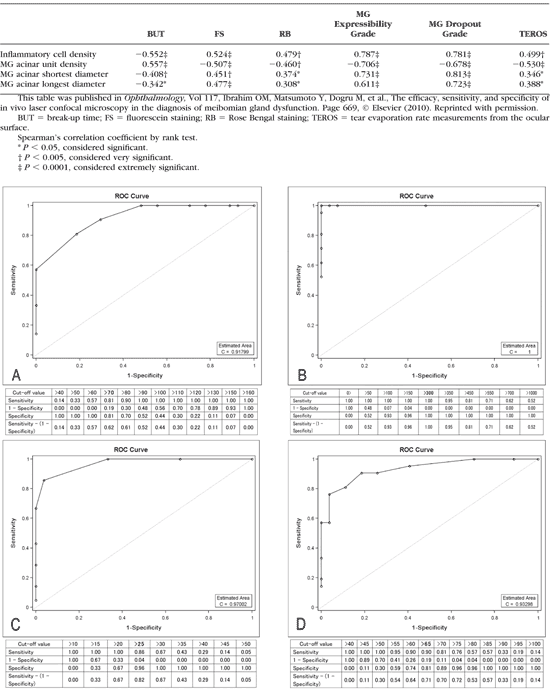
307. Ibrahim OM, Matsumoto Y, Dogru M, et al. The efficacy, sensitivity, and specificity of in vivo laser confocal microscopy in the diagnosis of meibomian gland dysfunction. Ophthalmology . 2010;117(4):665–672.
308. Chew CK, Jansweijer C, Tiffany JM, Dikstein S, Bron AJ. An instrument for quantifying meibomian lipid on the lid margin: the meibometer. Curr Eye Res. 1993;12:247–254.
309. Chew CK, Hykin PG, Jansweijer C, Dikstein S, Tiffany JM, Bron AJ. The casual level of meibomian lipids in humans. Curr Eye Res. 1993;12:255–259.
310. Yokoi N, Mossa F, Tiffany JM, Bron AJ. Assessment of meibomian gland function in dry eye using meibometry. Arch Ophthalmol. 1999;117:723–930.
311. Komuro A, Yokoi N, Kinoshita S, Tiffany JM, Bron AJ, Suzuki T. Assessment of meibomian gland function by a newly-developed laser meibometer. Adv Exp Med Biol. 2002;506:517–520.
312. Bron AJ, Tiffany JM, Gouveia SM, Yokoi N, Voon LW. Functional aspects of the tear film lipid layer. Exp Eye Res . 2004;78:347–360.
313. Doane MG. An instrument for in vivo tear film interferometry. Optom Vis Sci. 1989;66:383–388.
314. Danjo Y, Hamano T. Obeservation of precorneal tear film in patient with Sjogren's syndrome. Acta Ophthalmol Scand. 1995; 73:501–505.
315. Korb DR, Baron DF, Herman JP, et al. Tear film lipid layer thickness as a function of blinking. Cornea . 1994;13:354–359.
316. Guillon JP. Tear film photography and contact lens wear. J Br Contact Lens Assoc. 1982;5:84–87.
317. Goto E, Dogru M, Kojima T, Tsubota K. Computer-synthsis of an interference color chart of human tear lipid layer by a colorimetric approach. Invest Ophthalmol Vis Sci. 2003;44:4693–4697.
318. Goto E, Tseng SC. Differentiation of lipid tear deficiency dry eye by kinetic analysis of tear interference images. Arch Ophthalmol. 2003;121:173–180.
319. Goto E, Tseng SC. Kinetic analysis of tear interference images in aqueous tear deficiency dry eye before and after punctual occlusion. Inves t Ophthalmol Vis Sci. 2003;44:1897–1905.
320. Yokoi N, Takehisa Y, Kinoshita S. Correlation of tear lipid layer interference patterns with the diagnosis and severity of dry eye. Am J Ophthalmol. 1996;122:818–824.
321. King-Smith PE, Fink BA, Nichols JJ, Nichols KK, Hill RM. Interferometric imaging of the full thickness of the precorneal tear film. J Opt Soc Am A Opt Image Sci Vis . 2006;23(9):2097–2104.
322. Kimball SH, King-Smith PE , Nichols JJ. Evidence for the major contribution of evaporation to tear film thinning between blinks. Invest Ophthalmol Vis Sci. 2010;51:6294–6297.
323. King-Smith PE , Hinel EA, Nichols JJ. Application of a novel interferometric method to investigate the relation between lipid layer thickness and tear film thinning. Invest Ophthalmol Vis Sci. 2010;51(5):2418–2423.
324. Yokoi N, Yamada H, Mizukusa Y, et al. Rheology of tear film lipid layer spread in normal and aqueous tear-deficient dry eyes. Invest Ophthalmol Vis Sci. 2008;49:5319–5324.
325. Maruyama K, Yokoi N, Takamata A, Kinoshita S. Effect of environmental conditions on tear dynamics in soft contact lens wearers. 1: Invest Ophthalmol Vis Sci. 2004;45:2563–2568.
326. McCulley JP, Shine WE. Changing concepts in the diagnosis and management of blepharitis. Cornea. 2000;19:650–658.
327. Hisatake K, Tanaka S, Aizawa Y. Evaporation rate of water in a vessel. J Appl Phys. 1993; 11:7395–7401.
Mishima S, Maurice DM. The oily layer of the tear film and evaporation from the corneal surface. Exp Eye Res. 1961;1: 39–45.
329. Iwata S, Lemp M, Holly FJ, Dohlman CH. Evaporation rate of water from the pre-corneal tear film and cornea in the rabbit. Invest Ophthalmol. 1969;8:613–619.
330. Craig JP, Tomlinson A. Importance of the lipid layer in human tear film stability and evaporation. Optom Vis Sci. 1997;74:8–13.
331. Hamano H, Hori M, Kawabe H, et al. Modification of the superficial layer of the tear film by the secretion of the meibomian glands. Folio Ophthalmol Japonica. 1980;31:353–360.
332. Hamano H, Hori M, Mitsunaga S. Measurement of evaporation rate of water from theprecorneal tear film and contact lenses. Contacto. 1981;25:7–14.
333. Cedarstaff TH, Tomlinson A. A comparative study of tear evaporation rates and water content of soft contact lenses. Am J Optom Physiol Opt . 1983;60(3):167–174.
334. Trees G, Tomlinson A. Effect of artificial tear solutions and saline on tear film evaporation. Optom Vis Sci. 1990;67:886–890.
335. Rolando M, Refojo MF. Tear evaporimeter for measuring water evaporation rate from the tear film under controlled conditions in humans. Exp Eye Res. 1983;36:25–33.
336. Yamada M, Tsubota K. Measurement of tear evaporation from ocular surface (in Japanese). Nippon Ganka Zasshi. 1990;11: 1061–1070.
337. Mathers WD. Ocular evaporation in meibomian gland dysfunction and dry eye. Ophthalmology. 1993;100:347–351.
338. Goto E, Endo K, Suzuki A, et al. Tear evaporation dynamics in normal subjects and subjects with obstructive meibomian gland dysfunction. Invest Ophthalmol Vis Sci. 2003;44:533–539.
339. Mathers W. Evaporation from the ocular surface. Exp Eye Res. 2004;78:389–394.
340. Tomlinson A, Khanal S. Assessment of tear film dynamics: quantification approach. Ocul Surf. 2005;3:81–95.
341. Borchman D, Foulks GN, Yappert MC, Mathews J, Leake K, Bell J. Factors affecting evaporation rates of tear film components measured in vitro. Eye Contact Lens. 2009;35(1):32–37.
342. Nichols JJ, Mitchell GL. King-Smith PE. Thinning rate of the precorneal and prelens tear films. Invest Ophthalmol Vis Sci. 2005;46:2353–2361.
343. Tomlinson A, Pearce EI, Simmons PA, et al. Effect of oral contraceptives on tear physiology. Ophthalmic Physiol Opt. 2001;21: 9–16.
344. Mathers WD, Lane JA, Sutphin JE, Zimmerman MB. Model for ocular tear film function. Cornea. 1996;15:110–119.
345. Tsubota K, Yamada M. Tear evaporation from the ocular surface. Invest Ophthalmol Vis Sci. 1992;33:2942–2950.
346. Shimazaki J, Sakata M, Tsubota K. Ocular surface changes and discomfort in patients with meibomian gland dysfunction. Arch Ophthalmol. 1995;113:1266–1270.
347. McCulley JP, Shine WE, Aronowicz J, Oral D, Vargas J. Hyposecretory/hyperevaporative KCS: tear characteristics. Trans Am Ophthalmol Soc. 2003;101:141–154.
348. Khanal S, Tomlinson A, Diaper CJ. Tear physiology of aqueous deficiency and evaporative dry eye. Optom Vis Sci. 2009;86(11): 1235–1240.
349. Khanal S. Dry eye: diagnosis and management. PhD Thesis, Glasgow Caledonian University , 2006:110.
350. DEWS. Methodologies to diagnose and monitor dry eye disease: report of the Diagnostic Methodology Subcommittee of the International Dry Eye WorkShop (2007). Ocul Surf. 2007;5(2):108–152.
351. Tomlinson A, Giesbrecht C. Effect of age on human tear film evaporation in normals. Adv Exp Med Biol . 1994;350;271–274.
352. Mathers WD, Daley TE. Tear flow and evaporation in patients with and without dry eye. Ophthalmology. 1996;103:664–669.
353. Mathers WD, Binarao G, Petroll M. Ocular water evaporation and the dry eye: a new measuring device. Cornea. 1993;12:335–340.
354. Glass GV. Integrating findings: the meta-analysis of research. Rev Res Educ , 1977;5:351–379.
355. Tomlinson A, Doane MG, McFadyen A. Inputs and outputs of the lacrimal system: review of product and evaporative loss. Ocul Surf . 2009;7:186–198.
356. King-Smith E, Nichols JJ, Nichols KK, Fink BA, Braun RJ. Contributions of evaporation and other mechanisms to tear film thinning and break-up. Optom Vis Sci . 2008;85:623–630.
357. King-Smith E, Fink BA, Nichols JS, Nichols KK, Hill RM. Interferometric imaging of the full thickness of the precorneal tear film. J Opt Soc Am Opt Image Sci Vis 2006;23:2097–2104.
358. Kimball S, King-Smith E, Nichols JJ. Evidence for the major contribution of evaporation to tear film thinning between blinks. Invest Ophthalmol Vis Sci. 2010;51:5294–6297.
359. Shine WE, McCulley JR. Polar lipids in human meibomian gland secretions. Curr Eye Res , 2003;26(2):89–94.
360. Ham BM, Jacob JT, Keese MM, Cole RB. Identification, quantification and comparison of major non-polar lipids in normal and dry eye tear lipidomes by electrospray tandem mass spectrometry. J Mass Spectrom . 2004;39(11):1321–1336.
361. Butovich IA. On the lipid composition of human meibum and tears: Comparative analysis of nonpolar lipids. Invest Ophthalmol Vis Sci. 2008;49(9):3779–3789.
362. Borchman D, Foulk GN, Yappert MC, Tang D, Ho DV. Spectroscopic evaluation of human tear lipids. Chem Phys Lipids . 2007; 147(2):87–102.
363. Butovich IA, Millar TJ, Ham BM. Understanding and analyzing meibomian lipids: a review. Curr Eye Res . 2008;33(5–6):405–420.
364. Nichols KK, Ham BM, Nichols JJ, Ziegler C, Green-Church KB. Identification of fatty acids and fatty acid amides in human meibomian gland secretions. Invest Ophthalmol Vis Sci. 2007;48(1): 34–39.
365. Shine WE, McCulley JP. Meibomian gland triglyceride fatty acid differences in chronic blepharitis patients. Cornea . 1996;15(4) 340–346.
366. Ham BM, Cole RB, Jacob JT. Identification and comparison of the polar phospholipids in normal and dry eye rabbit tears by MALDITOF mass spectrometry. Invest Ophthalmol Vis Sci. 2006;47(8): 3330–3338.
367. Shine WE, McCulley JP. Meibomianitis: polar lipid abnormalities. Cornea . 2004;23(8):781–783.
368. Shine WE, McCulley JP. Keratoconjunctivitis sicca associated with meibomian secretion polar lipid abnormality. Arch Ophthalmol . 1998;116(7):849–852.
369. Joffre C, Souchier M, Gre´goire S, et al. Differences in meibomian fatty acid composition in patients with meibomian gland dysfunction and aqueous-deficient dry eye. Br J Ophthalmol . 2008;92(1): 116–119.
370. Norn MS. Tear secretion in normal eyes: estimated by a new method–-the lacrimal streak dilution test. Acta Ophthalmol. 1965;43:567–573.
371. Furukawa RE, Polse KA. Changes in tear flow accompanying ageing. Am J Optom Physiol Opt. 1978;55:69–74.
372. van Best JA, Oosterhuis JA. Computer fluorophotometry. Doc Ophthalmol. 1983;56;89–97.
373. Webber WRS, Jones DP, Wright P. Fluorophotometric measurements of tear turnover rate in normal healthy person: evidence for a circadian rhythm. Eye. 1987;1:615–620.
374. Occhipinti JR, Mosier MA, La Motte J, Monji GT. Fluorophotometric measurement of human tear turnover rate. Curr Eye Res . 1988;7:995–1000.
375. Gobbels M, Goebels G, Breitbach R, Spitznas M. Tear secretion in dry eyes as assessed by objective fluorophotometry. Ger J Ophthalmol. 1992;1:350–353.
376. van Best JA, Benilez Del Castillo JM, Coulangeon LM. Measurement of basal tear turnover using a standardized protocol: European Concerted Action on Ocular Fluorometry. Graefes Arch Clin Exp Ophthalmol. 1995;233:1–7.
377. Mishima S. Some physiological aspects of the precorneal tear film. Arch Ophthalmol. 1965;73:233–241.
378. Kuppens EV, Stolwijk TR, de Keizer RJW, aBest JA. Basal tear turnover and topical timolol in glaucoma patients and healthy controls by fluorophotometry. Invest Ophthalmol Vis Sci. 1992; 33:3442–3448.
Khanal S, Tomlinson A, Diaper CJ. Tear physiology of aqueous deficiency and evaporative dry eye. Optom Vis Sci. 2009;86(11): 1235–1240.
380. McCann LC, Tomlinson A, Pearce EI, Diaper C. Tear and meibomian gland function in blepharitis. Eye Contact Lens . 2009;35(4): 203–208.
381. van Best JA, Benitez del Castillo JM, Coulangeon LM. Measurement of basal tear turnover using a standardized protocol. European concerted action on ocular fluorometry. Graefes Arch Clin Exp Ophthalmol. 1995;233(1):1–7.
382. Scherz W, Doane MG, Dohlman CH. Tear volume in normal eyes and keratoconjunctivitis sicca. Albrecht Von Graefes Arch Klin Exp Ophthalmol . 1974;192(2):141–150.
383. Keijser S, van Best JA, van der Lelij A, Jager MJ. Reflex and steady state tears in patients with latent stromal herpetic keratitis. Invest Ophthalmol Vis Sci. 2002;43:87–91.
384. Mathers WD, Lane JA, Sutphin JE, Zimmerman MB. Model for ocular tear film function. Cornea. 1996;15:110–119.
385. Mathers WD, Daley TE. Tear flow and evaporation in patients with and without dry eye. Ophthalmology. 1996;103:664–669.
386. Tomlinson A, Doane MG, McFadyen A. Inputs and outputs of the lacrimal system: Meta-analysis of product and evaporative loss. Ocul Surf. 2009:7:17–29.
387. Holly FJ: Physical chemistry of the normal and disordered tear film. Trans Ophthalmol Soc UK . 1985;104:374–380.
388. Mainstone JC, Bruce AS, Golding TR. Tear meniscus measurement in the diagnosis of dry eye. Curr Eye Res. 1996;15:653–661.
389. Bron AJ: The Doyne Lecture: reflections on the tears. Eye. 1997; 11:583–602.
390. Yokoi N, Bron AJ, Tiffany JM, Brown NAP, Hsuan JD, Fowler CW. Reflective meniscometry: a non-invasive method to measure tear meniscus curvature. Br J Ophthalmol. 1999;83:92–97.
391. Yokoi N, Bron AJ, Tiffany JM, Kinoshita S. Reflective meniscometry: a new field of dry eye assessment. Cornea. 2000;19: S37–S43.
392. Wang J, Palakuru JR, Aquavella JV. Correlations among upper and lower tear menisci, non-invasive tear break-up time, and the Schirmer test. Am J Ophthalmol. 2008;145:795–800.
393. Yokoi N, Komuro A. Non-invasive methods of assessing the tear film. Exp Eye Res 2004;78:399–407.
394. Oguz H, Yokoi N, Kinoshita S. The height and radius of the tear meniscus and methods for examining these parameters. Cornea. 2000;19:497–500.
395. Farrell J, Patel S, Grierson DG, Sturrock RD. A clinical procedure to predict the value of temporary occlusion therapy in keratoconjunctivitis scicca. Ophthalmic Physiol Opt. 2003;23:1–8.
396. Yokoi N, Bron AJ, Tiffany JM, Maruyama K, Komuro A, Kinoshita S. Relationship between tear volume and tear meniscus curvature. Arch Ophthalmol. 2004;122:1265–1269.
397. Gilbard JP, Farris RL, Santamaria J 2nd. Osmolarity of tear microvolumes in keratoconjunctivitis sicca. Arch Ophthalmol. 1978; 96(4):677–681.
398. Farris RL. Tear osmolarity–a new gold standard? Adv Exp Med Biol. 1994;350:495–503.
399. Gilbard JP, Dartt DA. Changes in rabbit lacrimal gland fluid osmolarity with flow rate. Invest Ophthalmol Vis Sci. 1982;23(6): 804–806.
400. Sullivan BD. Clinical resorts of a first generation laboratory-onchip nanolitre tear film osmometer. Ocul Surf. 2005;3:S31.
401. Nelson JD, Wright JC. Tear film osmolality determination: an evaluation of potential errors in measurement. Curr Eye Res . 1986;5(9):677–681.
402. Terry JE, Hill RM. Human tear osmotic pressure: diurnal variations and the closed eye. Arch Ophthalmol. 1978;96(1):120–122.
403. White KM, Benjamin WJ, Hill RM. Human basic tear fluid osmolality. I. Importance of sample collection strategy. Acta Ophthalmol ( Copenh ). 1993;71(4):524–529.
404. Tomlinson A, McCann LC, Pearce EI. Comparison of human tear film osmolarity measured by electrical impedance and freezing point depression techniques. Cornea. 2010(9):1036–1041.
405. Tomlinson A, Khanal S, Ramaesh K, et al. Tear film osmolarity: determination of a referent for dry eye diagnosis. Invest Ophthalmol Vis Sci. 2006;47(10):4309–4315.
406. Khanal S, Tomlinson A, Diaper CJ. Tear physiology of aqueous deficiency and evaporative dry eye. Optom Vis Sci. 2009;86(11): 1235–1240.
407. Sullivan BD, Whitmer D, Nichols KK, et al. An objective approach to dry eye disease severity. Invest Ophthalmol Vis Sci. 2010;51: 6125–6130.
408. Tomlinson A, Doane MG, McFadyen A. Inputs and outputs of the lacrimal system: review of production and evaporative loss. Ocul Surf. 2009;(4):186–198.
409. Tomlinson A, Khanal S. Assessment of tear film dynamics: quantification approach. Ocul Surf. 2005;3:81–95.
410. Xu KP, Yagi Y, Toda I, Tsubota K. Tear function index: a new measure of dry eye. Arch Ophthalmol. 1995;113:84–88.
411. Xu K, Tsubota K. Correlation of tear clearance rate and fluorophotometric assessment of tear turnover. Br J Ophthalmol. 1995; 79:1042–1045.
412. Tsubota K. Tear dynamics and dry eye. Prog Retin Eye Res. 1998;17:565–596.
413. Mathers WD, Choi D. Cluster analysis of patients with ocular surface disease, blepharitis and dry eye. Arch Ophthalmol. 2004; 122:1700–1704.
414. McCann LC, Tomlinson A, Pearce EI, Kaye SB, Fisher AC. A clinical alternative to fluorophotometry for measuring tear production in the diagnosis of dry eye. Cornea . 2010;29:745–750.
415. Mathers WD. Why the eye becomes dry: a cornea and lacrimal gland feedback model. CLAO J. 2000;26:159–165.
416. Sahlin S, Chen E. Evaluation of the lacrimal drainage function by the drop test. Am J Ophthalmol. 1996;122:701–708.
417. Tomlinson A, Doane MG, McFadyen A. Inputs and outputs of the lacrimal system: review of production and evaporative loss. Ocul Surf. 2009(4):186–198.
418. Mathers WD, Lane JA, Sutphin JE, Zimmerman MB. Model for ocular tear film function. Cornea. 1996;15:110–119.
419. Craig JP, Tomlinson A. Importance of the lipid layer in human tear film stability and evaporation. Optom Vis Sci. 1997;74:8–13.
420. Levin MH, Verkman AS. Aquaporin-dependent water permeation at the mouse ocular surface: in vivo microfluorimetric measurements in cornea and conjunctiva. Invest Ophthalmol Vis Sci. 2004;45(12):4423–4432.
421. King-Smith PE, Fink BA, Nichols JJ, Nichols KK, Braun RJ, McFadden GB. The contribution of lipid layer movement to tear film thinning and breakup. Invest Ophthalmol Vis Sci. 2009; 50(6):2747–2756. |